CFP™ Fluorophores
The Cleavable TSA Fluorophores
How does the cleavable TSA fluorophore work?
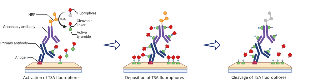
Similar to tyramide signal amplification (TSA), CFP cleavable TSA fluorophores leverage the catalytic activity of horseradish peroxidase (HRP) to enable the covalent deposition and binding of labeled fluorophores onto target proteins or nucleic acid sequences in situ. Our fluorophores are engineered for much higher reactivity than tyramide fluorophores, making the imaging process significantly faster and more robust than conventional TSA-based applications. Unlike conventional TSA fluorophores, CFP fluorescent signals can be cleaved using mild cleavage reagents, enabling ultra-sensitive reiterative staining with standard antibodies. Furthermore, CFP Fluor technology is highly versatile and can be seamlessly integrated into protocols requiring HRP-based detection, including immunohistochemistry (IHC), immunocytochemistry (ICC), immunofluorescence (IF), in situ hybridization (ISH), and proximity ligation assays (PLA).
Choices of CFP Fluorophores
Key Features of CFP Fluor:
- Ultra-sensitive detection of low-abundance targets
- Conserve precious antibodies. CFP fluorophores achieve equivalent levels of sensitivity with a significant reduction in primary antibody
- Compatible with other fluorescent markers, detection techniques making it suitable for co-detection applications
- The cleavable TSA fluorophores unlock the capability of cyclic staining for high-multiplex multiomics spatial applications
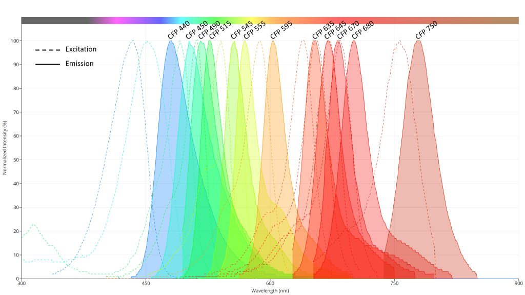
CFP™ Fluorophore Spectral Reference Table
| CFP Fluorophores | Equivalent Fluorophores | Excitation (peak nm) | Emission (peak nm) |
|---|---|---|---|
| CFP 440 | Alexa 440 | 435 | 480 |
| CFP 450 | Opal 480 | 450 | 502 |
| CFP 490 | Opal 520 | 490 | 515 |
| CFP 515 | Opal 540 | 510 | 527 |
| CFP 545 | Alexa 546 | 540 | 557 |
| CFP 555 | Opal 570 | 557 | 570 |
| CFP 595 | Opal 620 | 587 | 603 |
| CFP 635 | Opal 650 | 640 | 655 |
| CFP 645 | Alexa 647 | 655 | 670 |
| CFP 670 | Opal 690 | 670 | 682 |
| CFP 680 | Alexa 680 | 685 | 701 |
| CFP 750 | Opal 780 | 757 | 780 |